An electrocardiogram (EKG or ECG) is a non-invasive medical test that records the electrical activity of the heart. It is a crucial diagnostic tool used to detect heart problems, monitor heart health, and evaluate the effectiveness of treatments. The procedure involves placing electrodes on the skin to capture the heart’s electrical signals and display them […]
What is it?
An electrocardiogram (EKG or ECG) is a test that measures the electrical activity of the heart over a period of time. It is used to diagnose various heart conditions, including arrhythmias, heart attacks, and other cardiac disorders. The test provides a visual representation of the heart’s electrical impulses, showing the timing and duration of each phase of the heartbeat.
What happens
during the procedure:
During an EKG procedure, the following steps typically occur:
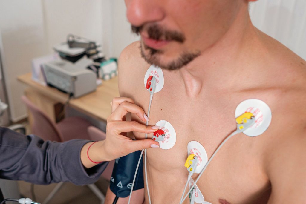
Preparation: The patient is asked to remove any jewelry and upper body clothing. The skin is cleaned to ensure good electrode contact.
Electrode Placement: Small adhesive electrodes are placed on the chest, arms, and legs. Typically, 10 electrodes are used to record the heart’s activity from multiple angles.
Recording: The electrodes are connected to the EKG machine, which records the electrical signals of the heart. The patient is asked to lie still and breathe normally while the recording is made. The test usually takes a few minutes.
Monitoring: The EKG machine displays the heart’s electrical activity as a series of waveforms on a monitor or prints them on paper.
Completion: The electrodes are removed, and the patient can resume normal activities. The recorded data is then analyzed by a healthcare provider.
Benefits:
Non-Invasive: The procedure is painless and does not require any surgical intervention.
Quick Results: Provides immediate results that can be used for prompt diagnosis and treatment.
Diagnostic Accuracy: Helps detect a wide range of heart conditions, including arrhythmias, myocardial infarctions, and electrolyte imbalances.
Monitoring: Useful for ongoing monitoring of patients with known heart conditions or those undergoing treatment.
Baseline Data: Establishes baseline heart activity for comparison with future EKGs.
Things to keep in mind:
Movement: Avoid movement during the test to prevent artifacts (distortions) in the recordings.
Electrode Placement: Proper placement of electrodes is crucial for accurate readings; inform the technician of any skin issues or discomfort.
Medications: Inform the healthcare provider about any medications being taken, as some can affect heart activity.
Follow-up: Additional tests may be required if the EKG shows abnormal results to confirm a diagnosis or evaluate the severity of a condition.
Limitations: An EKG provides information about the heart’s electrical activity but does not show the structure of the heart. Further imaging tests may be needed for structural information.
Alternatives:
Holter Monitor: A portable device worn for 24-48 hours to continuously record heart activity over an extended period.
Event Monitor: Similar to a Holter monitor, but used for longer periods (weeks to months) and records heart activity only when triggered by the patient or automatically during abnormal rhythms.
Echocardiogram: Uses ultrasound waves to create images of the heart’s structure and function.
Stress Test: Monitors heart activity during physical exercise to detect issues not apparent at rest.
Cardiac MRI: Provides detailed images of the heart’s structure and function using magnetic resonance imaging.
Blood Tests: Measure levels of certain biomarkers that indicate heart damage or stress, such as troponins.